Cardiac CT
Pathology Testing Labs Specialists in Mumbai
Cardiac CT:
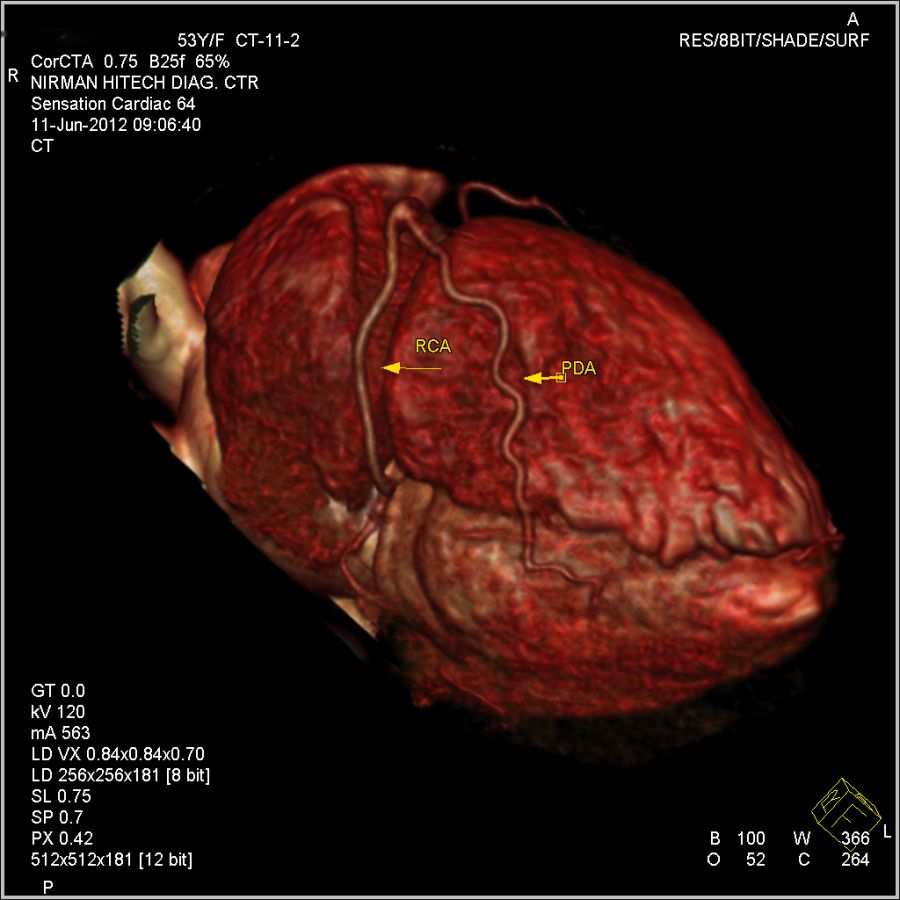
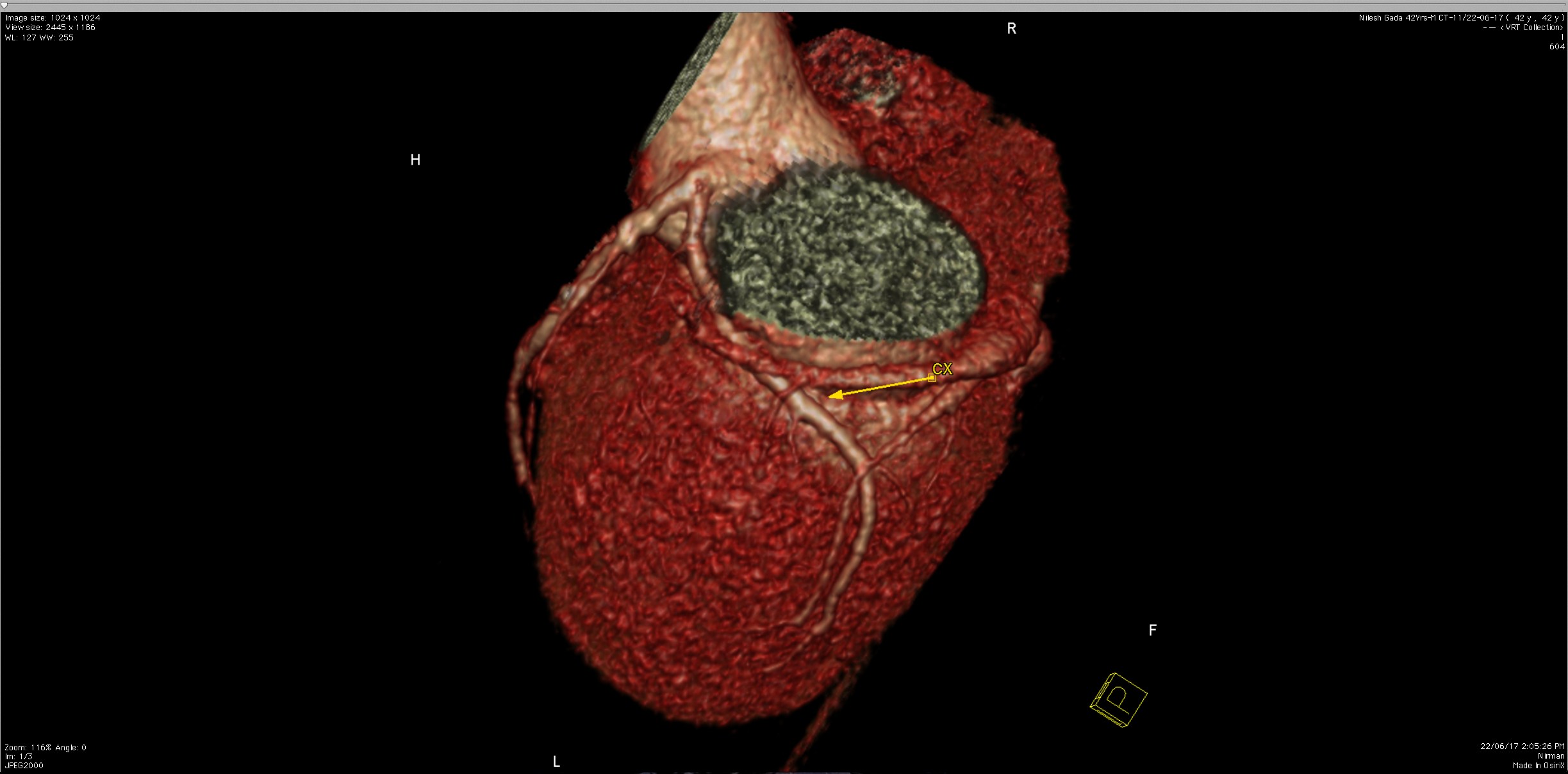
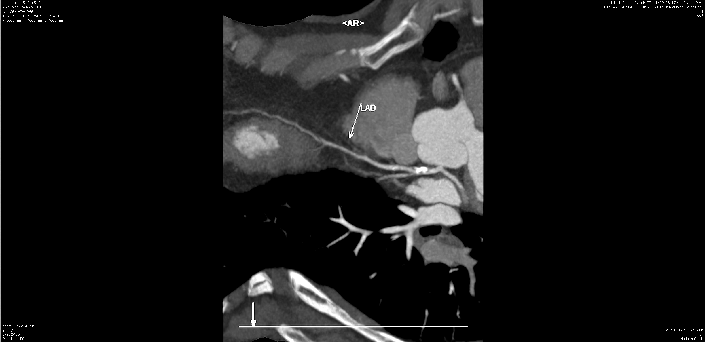
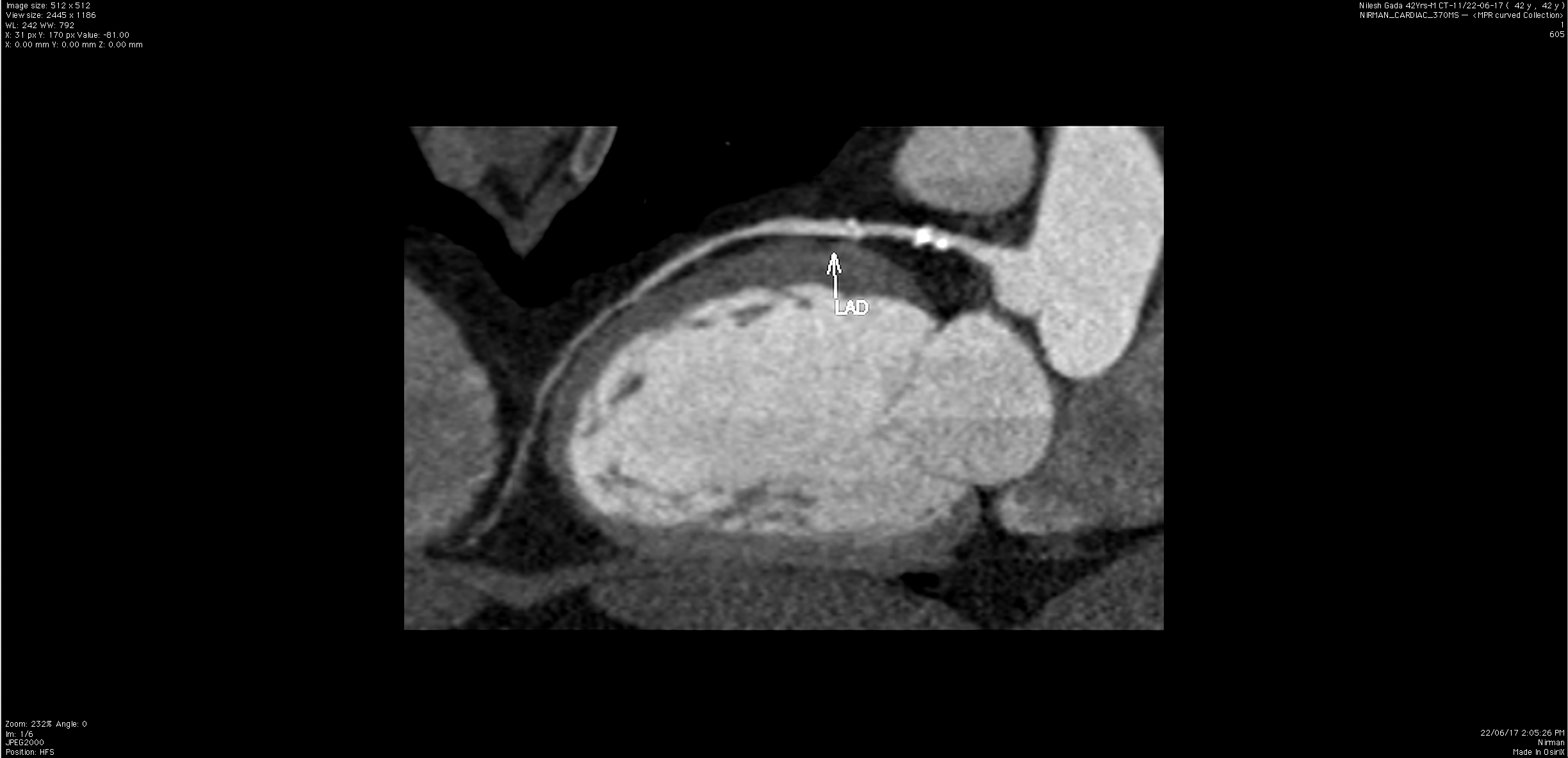
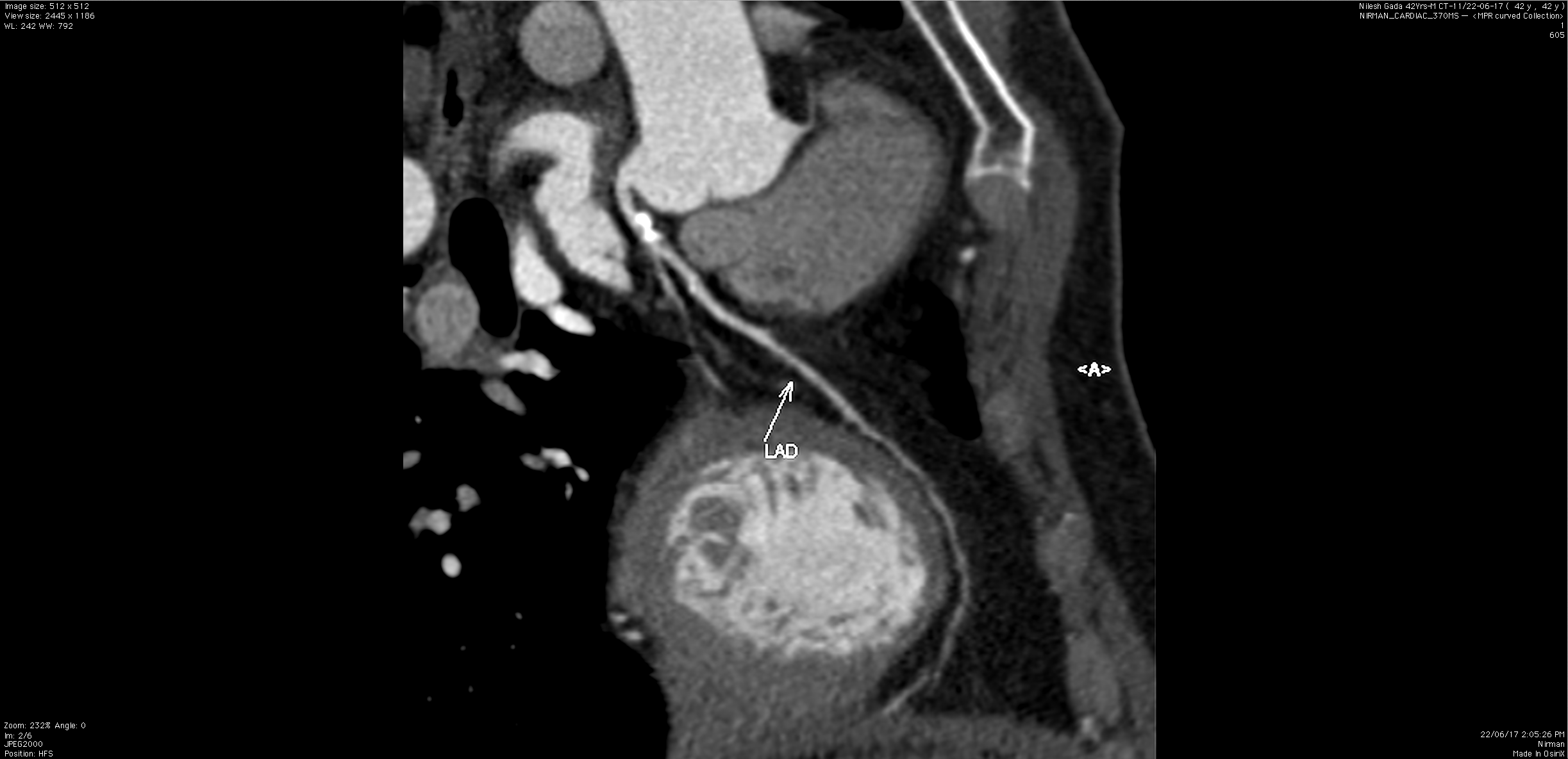
CT scanning of the heart (CT coronary angiogram) is a procedure used to assess the extent of occlusion in the coronary arteries, usually in order to diagnose coronary artery disease. The patient is injected with an intravenous dye (iodine) and then their heart is scanned using a high speed CT scanner, allowing radiographers to assess the blood flow to their heart muscle During this procedure we do a plain scan to check the calcium content of the coronary arteries and then we perform a non invasive CT coronary angiography to assess the coronary lumen.
What is the Need For CARDIAC CT?
Coronary Artery disease (CAD) is the leading cause of death in the industrialized world. 50% of patients first present with "sudden death". However there are classified risk factors for developing CAD. Thus risk assessment and early diagnosis or "screening" are widely accepted strategies to combat CAD. Even in high risk patients, if CAD is diagnosed at the early stage, the changes are usually reversible and the patient can go back to a low risk state, with suitable cardiologists follow up, and dietetic & lifestyle modification. Thus the interest in non-invasive modalities to diagnose CAD is growing in the medical world. Cardiac CT provides just the tools physicians need by offering non-invasive visualization of the coronary artery lumen and plaques in the vessel wall as well as measurement of the coronary calcium burden.
Thus We Offer:
Calcium scoring - for calcium content evaluation of the coronary arteries.
Coronary Artery Lumen evaluation
Argus Function evaluation of the ventricular muscle mass.
What Are The Current Applications of CT Coronary Angiography?
- Early detection of stenosis of the coronary artery lumen.
- Non- symptomatic high-risk patients.
- Risk factors include hypertension, diabetes, stroke, lipid disorders.
- Family history of the same.
- Smoking.
- High Stress.
- Exclusion of stenosis
- Atypical (unstable) chest pain.
- Refractory chest pain with doubtful coronary origin.
- Non-conclusive stress tests.
- Detection and / or exclusion of stenosis
- high risk patients.
- Prior to major (non-cardiac) Surgery.
- As a substitute to conventional coronary angiography
- Prior to percatareous coronary intervention.
- High risk patient like aortic disease.
- Adjuvant to coronary Angio
- Plaque characterization.
- Complicated coronary intubation.
- Total coronary occlusion.
- Follow-up in
- Percutaneous coronary intervention.
- Bypass Surgery.
- Evaluation of coronary anomalies.
- Evaluation of chest pain at emergency department.
- Evaluation of lifestyle, dietary or pharmacological interventions on progression /regression of coronary atherosclerosis.
What is Cardiac CT for Calcium Scoring?
Computed tomography, also known as CT or CAT scanning, uses a special machine to obtain multiple x-ray images of any part of the body. The images are much more detailed than those provided by conventional x-rays. In addition, CT can display many different types of tissue including blood vessels. Modern scanners use a technique called spiral or helical CT to obtain images from many angles. Computerized processing of these images creates cross-sections, or slices, of the area of interest. The images can then be examined on a computer monitor or printed out. Cardiac CT for calcium scoring is a non-invasive way of obtaining information about the location and extent of calcified plaque in the coronary arteries-the vessels that supply oxygen-containing blood to the heart wall. Plaque is a build-up of fat and other substances, including calcium, which in time can narrow the arteries or even close off blood flow to the heart. The result may be painful angina in the chest or a heart attack. Calcium is a marker of coronary artery disease. The findings on cardiac CT, expressed as a calcium score, may help decide what measures can be taken to avoid these events. Another name for this test is coronary artery calcium scoring.
How should I prepare for the procedure?
No special preparation is necessary in advance of a cardiac computed tomography (CT) examination. You may continue to take your usual medications, but should avoid caffeine and smoking for four hours before the exam. At the time of the exam you will be asked to disrobe above the waist, put on a gown, and remove any jewelry that could interfere with the CT scan. If your heart rate is 90 beats a minute or higher, you may be given a drug to slow the rate in order to obtain accurate CT images.
How does the procedure work?
During a computed tomography (CT) scan, the rotating gantry will emit x-rays that pass through the part of the body being examined-in this case the heart and coronary arteries. In spiral or helical CT, the patient passes through the scanner as the gantry rotates. Multiple detectors mounted on the gantry along with the x-ray tube record the radiation leaving the body. The result is that the x-ray beam follows a spiral path. The recorded images are reconstructed by computer using a special software program. Recently developed spiral CT scanners produce high-quality images in less than 10 seconds. This is especially important for elderly patients and those who cannot hold their breath for the required time. A negative cardiac CT scan that shows no calcification within the coronary arteries suggests that atherosclerotic plaque is minimal at most, and that the chance of coronary artery disease developing over the next two to five years is very low. A positive test means that coronary artery disease is present even if you have no symptoms. The amount of calcification-expressed as a score-may help to predict the likelihood of a myocardial infarction (heart attack) in the coming years
| Calcium Score | Presence of Plaque |
|---|---|
| 0 | No evidence of plaque |
| 1-10 | Minimal evidence of plaque |
| 11-140 | Mild evidence of plaque |
| 101-400 | Moderate evidence of plaque |
| Over 400 | Extensive evidence of plaque |
What Are The
Benefits vs. Risks?
Benefits
- Cardiac computed tomography (CT) for calcium scoring is a convenient and noninvasive way of evaluating the coronary arteries.
- The calcium score gives an idea of whether coronary artery disease (CAD) is present despite a lack of symptoms, or is likely to develop in the next few years.
- Cardiac CT takes little time and causes no pain.
- The exam does not require injection of contrast material and therefore avoids its possible side effects.
- The examination can suggest the presence of CAD even when the coronary arteries are less than 50 percent narrowed. Standard cardiac tests will not reliably detect this level of blockage, and more than half of all heart attacks occur with less than 50 percent narrowing.
Risks
- The exam exposes the patient to a limited amount of radiation. The dose is similar to that from 10 chest x-rays and about 10-20 percent of that received during a diagnostic cardiac catheterization procedure.
- Women should always inform their doctor, x-ray technologist or nurse if there is any possibility that they are pregnant.
- Cardiac CT sometimes is positive even though there is no significant blockage of the coronary arteries. As a result, the patient may undergo further tests that are not necessary and these tests might cause side effects.
 Interventional Radiology